

Medical Image and Signal Processing
Connecting imaging software, detector technology, neuroscience and preclinical research.
MEDISIP is a research group in the Department of Electronics and Information Systems (ELIS) at the Faculty of Engineering and Architecture, Ghent University. Its research spans medical imaging and signal processing, from software and hardware development to neuroscience and small-animal imaging.
Research & facilities


Explore MEDISIP in detail
Choose a section, then open a project or facility for its full description, figures and references.
The library preserves information from MEDISIP’s existing research pages, including earlier projects and publications.
Software lab
The Software lab mainly focusses on PET, SPECT, MRI and CT imaging. We have gained experience in advanced image reconstruction, system simulation and multimodal image analysis.
Current research
- Artificial Intelligence for medical image analysis and computer-aided brain tumour diagnosis
- Deep learning for positron emission tomography
- Internal radiotherapy dosimetry
- Positron range and acollinearity modeling for hybrid PET/CT and PET/MR imaging
- Quantitative imaging for radionuclide therapy
Past research
- Brain SPECT Imaging
- CT/MR phantoms for quality assessment
- Innovative Collimator for Compact Stationary SPECT
- Iterative CT reconstruction
- Joint reconstruction of activity and attenuation in listmode TOF PET
- Partial volume effect correction in PET imaging
- PET attenuation correction with a supplemental transmission source
- PET image reconstruction software QETIR
- PET system simulation
- PET/MRI attenuation correction
- Quantitative conductivity and permittivity imaging using magnetic resonance electrical properties tomography (MREPT)
- Quantitative image analysis for automated brain tumour analysis
- SPECT System Modeling and Simulations
- SPECT/MRI compatibility
- The new paediatric TOF-PET: a simulation study
Artificial Intelligence for medical image analysis and computer-aided brain tumour diagnosis
The increasing amount of data recorded and stored into electronic health records becomes overwhelming for healthcare professionals. This project aims to address the growing need to assist physicians in processing these large amounts of data through the use of Artificial Intelligence (AI). Especially in diagnostic imaging, AI can help radiologists meet the rising demand for imaging examinations and prevent diagnostic errors. In this project we will focus on designing AI for accurate and automatic brain tumour characterisation based on medical imaging.
In Belgium, 800 people are diagnosed with a primary brain tumour each year. Their life expectancy depends strongly on the tumour type and molecular characteristics, and ranges from several decades for low-grade tumours to only about one year for the most malignant types. An accurate diagnosis and prediction of genetic markers is therefore of primary importance. In this project, we will implement a method for automated, non-invase brain tumour characterisation based on pre-therapy MRI, using Convolutional Neural Networks.

Contact: Milan Decuyper
Deep learning for positron emission tomography
Positron emission tomography (PET) is commonly used for the detection and diagnosis of cancer. It works by injecting the patient with a positron-emitting radioactive tracer that gathers around cancerous tissue. The emitted positrons annihilate with nearby electrons, producing two back-to-back gamma photons. Scintillation crystals absorb these gamma photons and re-emit them as visible light, which is then detected by silicon photomultipliers (SiPMs). This information can then be used to reconstruct the 3D tracer distribution.
This project focuses on improving the overall performance of PET scanners by substituting or enhancing conventionally used signal processing techniques with modern deep learning algorithms. These algorithms are implemented both at the system level (for image reconstruction and denoising) and the detector level (for positioning and timing of the gamma absorptions). The improved scan quality allows for detection of diseases during earlier stages.
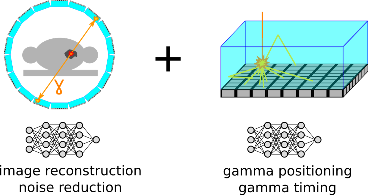
Contact: Jens Maebe and Milan Decuyper
Internal radiotherapy dosimetry
Internal vectorised radiotherapy are promising therapeutic modalities, increasingly used in the treatment of unresectable tumors. Optimization of the therapeutic outcome by maximizing the dose delivered to the tumor, while keeping the dose delivered to organs at risk at acceptable levels is the main goal of dosimetry. Systematically implemented dosimetry is essential in order to fully exploit the therapeutic potential of internal vectorised radiotherapy. Furthermore, accurate and precise dosimetric estimations are crucial for establishing a correlation between absorbed dose and treatment efficacy and safety. However, accuracy and precision in dosimetry may be hampered by potential systematic and stochastic errors respectively. The aim of this project is to assess the potential errors committed on the dosimetric calculations and to evaluate their biological impact on tumor dose-response and organ dose-toxicity correlation.
Contact: Gwennaëlle Marin
Positron range and acollinearity modeling for hybrid PET/CT and PET/MR imaging
The development of integrated devices to combine Positron Emission Tomography (PET) and Magnetic Resonance (MR) imaging has been a constant trend in multimodal imaging for the last years. However, a number of approaches have shown that positron range and the acollinearity are fundamental limitations of PET image resolution. According to these studies, its effect is especially relevant with some radionuclides currently used in clinical and preclinical studies such as 82Rb, 124I and 68Ga. The aim of this project is threefold: 1 – Modeling the positron range and acollinearity of clinically relevant PET isotopes for different tissue types with and without the presence of a magnetic field. 2 – Validation of the positron model using phantom measurements with different isotopes and (pre)clinical PET/MRI and PET/CT systems. 3 – Use of the attenuation map derived from CT, MRI or PET only data to account for tissue dependent positron range during reconstruction.
Contact: Paulo Caribé
Quantitative imaging for radionuclide therapy.
The improvement of the reconstruction method to make images more quantitative for the labeled microspheres. This will be done with phantom studies and using acquired clinical studies.
Contact : Karnsinee yasamut
Brain SPECT Imaging
Single Photon Emission Computed Tomography (SPECT) is a nuclear imaging technique often used for functional cerebral imaging. Current whole-body systems have shown to be clinically useful in a wide range of neurological diseases like dementia, cerebrovascular diseases and epilepsy. However, a dedicated brain SPECT system with a better sensitivity and resolution could result in better diagnosis and more effective treatment. A SPECT device consists of two main components: a detector and a collimator. We focus on innovative collimator design and optimized a multi-pinhole collimator insert for an existing ring of LaBr3 detectors.
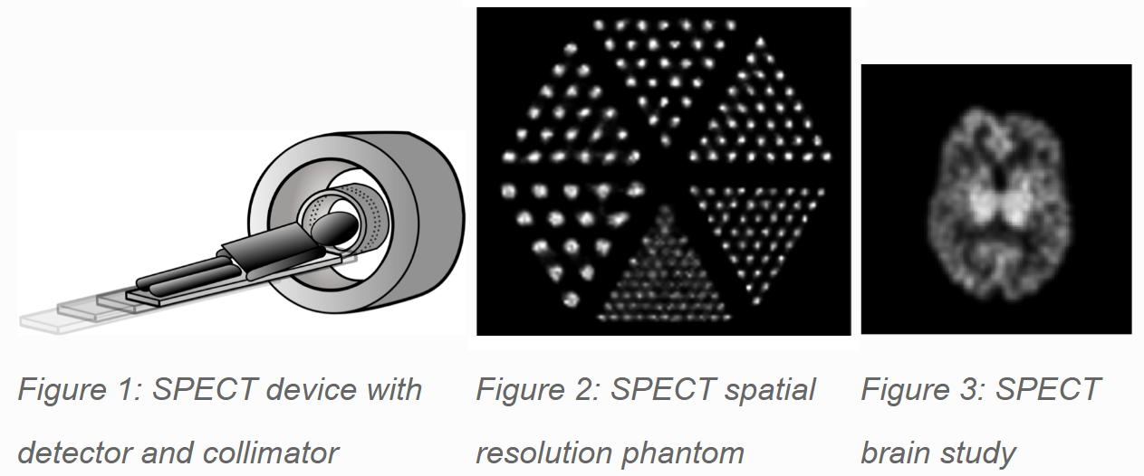
Researcher: Karen Van Audenhaege
CT/MR phantoms for quality assessment
Magnetic Resonance Imaging (MRI) is a radiation-free imaging technique. Therefore, when possible, should be preferred over other imaging modalities. However, Computed Tomography (CT) is still the gold standard in some organ imaging (lung, bone..) more challenging for MRI, and new MRI techniques should be validated against CT. We investigate the development of realistic CT/MR phantoms for quality assessment.
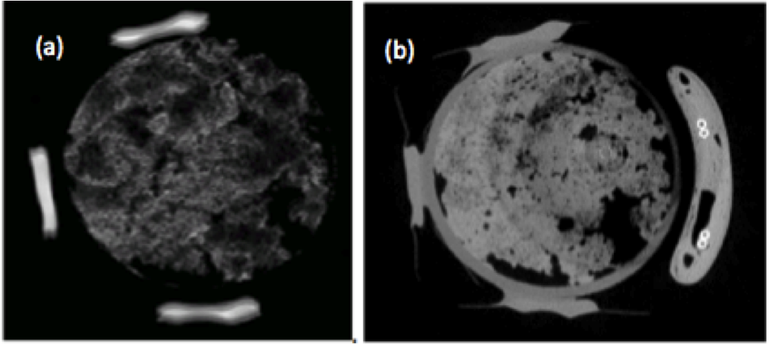
Researcher: Shandra Gutierrez Diaz.
Innovative Collimator for Compact Stationary SPECT
In Single Photon Emission Computed Tomography (SPECT) scanners, collimators are used in front of the detectors to help determine the direction from which gamma-rays originate. In order to reconstruct a 3D image of the radioisotope distribution, we need to measure radiation coming from the patient at different angles, and for this there are two main strategies: rotating parallel-hole collimators and stationary pinhole collimators.
We came up with and developed a new collimator concept, that marries the two approaches: a stationary parallel-hole collimator. Because it is stationary, it should be considerably cheaper, less time-consuming and provide better quality images than non-stationary SPECT scanners. On the other hand, because of its innovative design based on parallel-holes, we can image the same volume as other currently available stationary SPECT systems using a much more compact scanner, therefore saving both space and material costs.
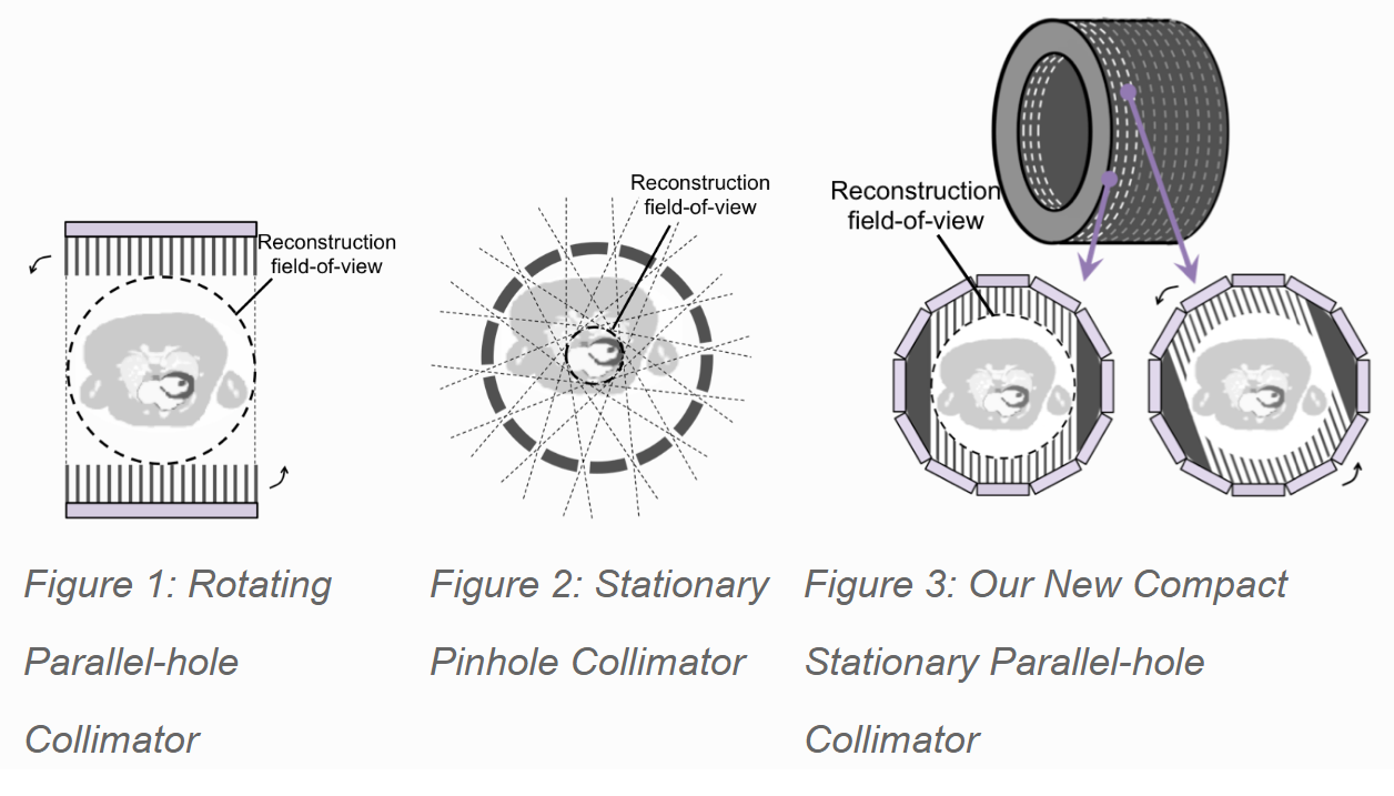
Researcher: Lara Pato
Iterative CT reconstruction
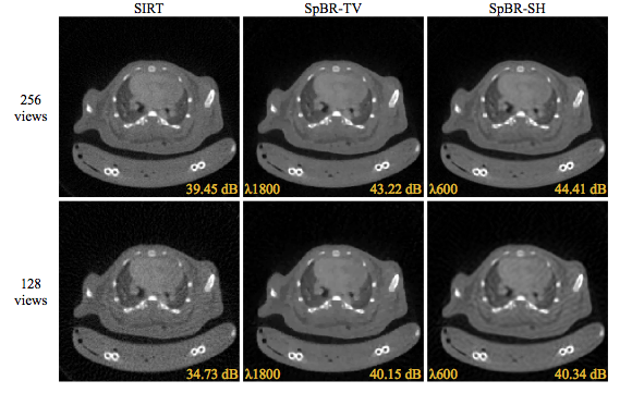
Reducing the image noise in in-vivo micro Computerized Tomography (μCT) is important to improve the image quality and maintain diagnostic confidence. Theoretically, the images can be improved by using longer acquisition times. However, because of the limitations on administrated dose and anesthetics other approaches need to be considered. The goal of this work is to find regularization for sparse-view reconstruction and image denoising in general.
Contact: Bert Vandeghinste
Joint reconstruction of activity and attenuation in listmode TOF PET
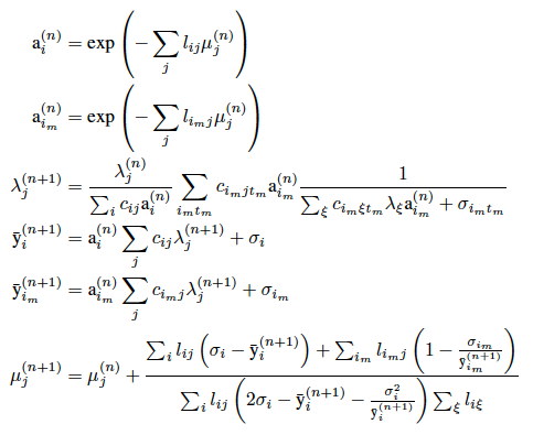
Joint reconstruction algorithms such as MLAA are capable to reconstruct both a PET image and the attenuation correction values (AC values) from Time-of-Flight PET data only. Although the inverse problem is ill-posed, the redundancy of the Time-of-Flight data allows to find AC values up to a constant scale factor. Current Medisip research focusses on listmode implementation of these algorithms and their extension to the case of prior information and to the case with background (scatter and random events).
Researcher: Ester D’Hoe
Partial volume effect correction in PET imaging
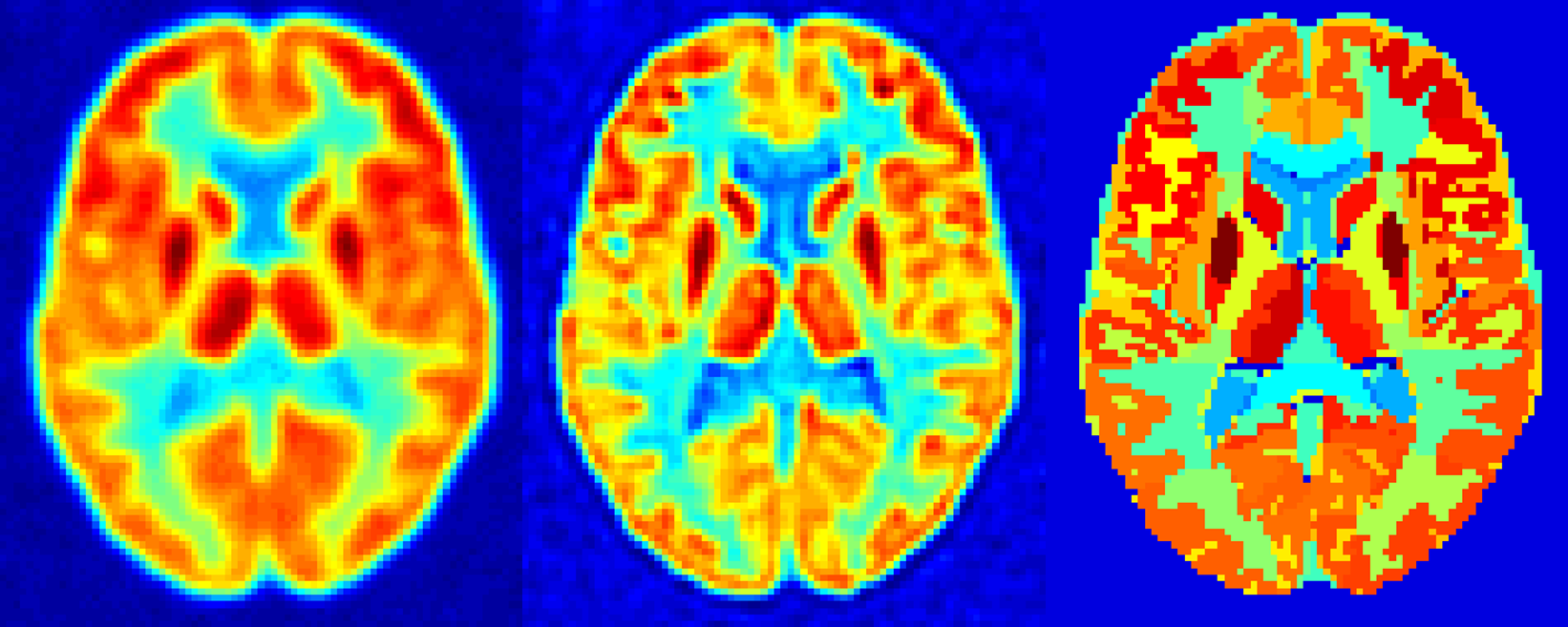
The partial volume effect is the deterioration of the image quality in a medical scan due to the limited resolution of the imaging system and the sampling of the image in discrete voxels. This results in a blurring of the image, and small structures showing a lower activity. Our group implements and develops post-reconstruction correction methods for this effect. This is done using information from the PET-scan itself (iterative deconvolution), or using anatomical information from a coregistered MRI-scan.
Researcher: Stijn Bonte
PET attenuation correction with a supplemental transmission source
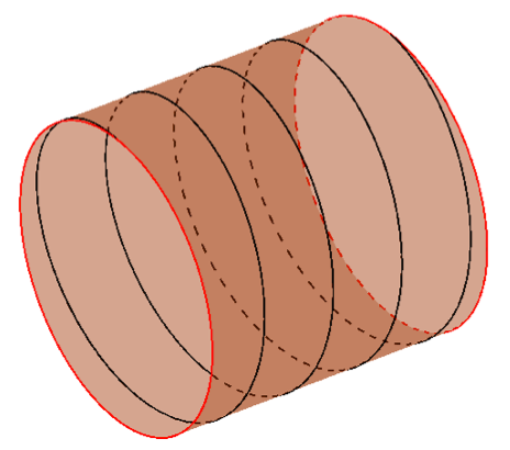
To obtain quantitative PET images, on needs to correct for the number of photons that were absorbed in the patient’s body. This so-called attenuation correction (AC) is in clinical settings mostly done by subsequently acquiring a CT scan. A simple bilinear scaling can convert CT Hounsfield units into PET linear attenuation coefficients. However, recently PET/MRI scanners have gained popularity while there exists no such relation between MR intensities and photon attenuation. Therefore, MR-based attenuation correction (MRAC) is a lot harder and not yet optimized for whole body PET imaging. The addition of a transmission source with a PET tracer can aid to find the attenuation correction values while keeping the dose to the patient low.
The research at Medisip lab focuses on the minimally required design for a transmission source. This is investigated in a theoretical way as well as with computer simulations. Future research will include real measurements with the theoretically optimal design.
Researcher: Ester D’Hoe
PET image reconstruction software QETIR
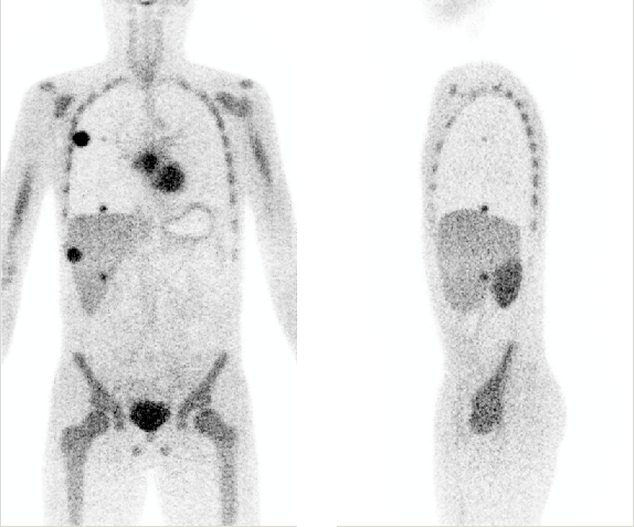
QETIR, which stands for Quantitative Emission Tomography Iterative Reconstruction is image reconstruction software for PET developed at Medisip. It utilizes listmode reconstruction as opposed to the more commonly used sinogram reconstruction. Implemented algorithms include (Time-of-Flight) MLEM, OSEM, MLTR, MLAA and MLAA+. Data from any scanner can be reconstructed if the scanner geometry is provided. Researchers interested in QETIR software can contact MEDISIP lab.
Researcher: Ester D’Hoe
PET system simulation
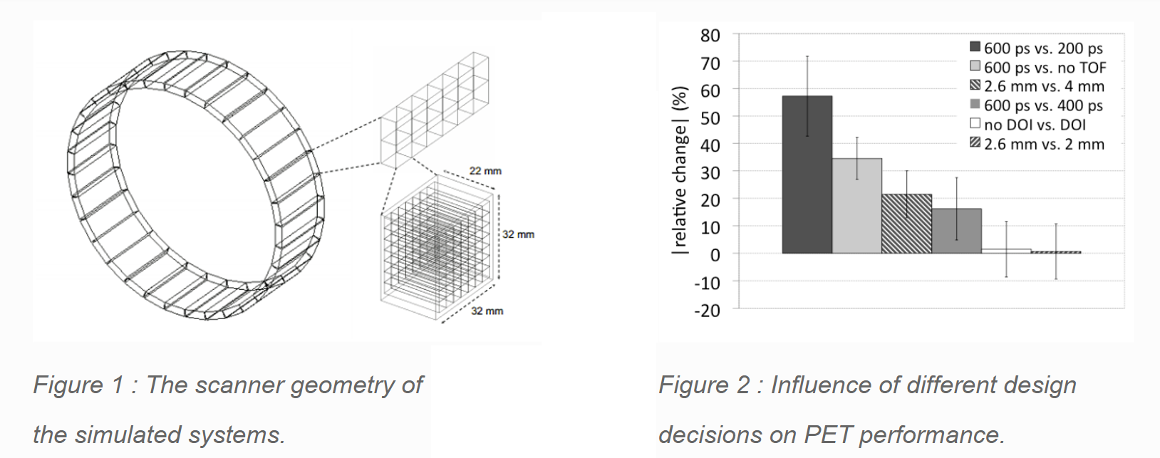
System simulations can help to determine the influence of PET design parameters such as spatial resolution, TOF resolution, energy resolution, and detection efficiency. In this project the simulations are performed using GATE, a standard Monte Carlo simulation tool for emission tomography. In particular the performance of the PET system suitable to operate in an MR environment is investigated.
Researchers: Pieter Mollet, Ester D’Hoe, Ekaterina Mikhaylova.
PET/MRI attenuation correction
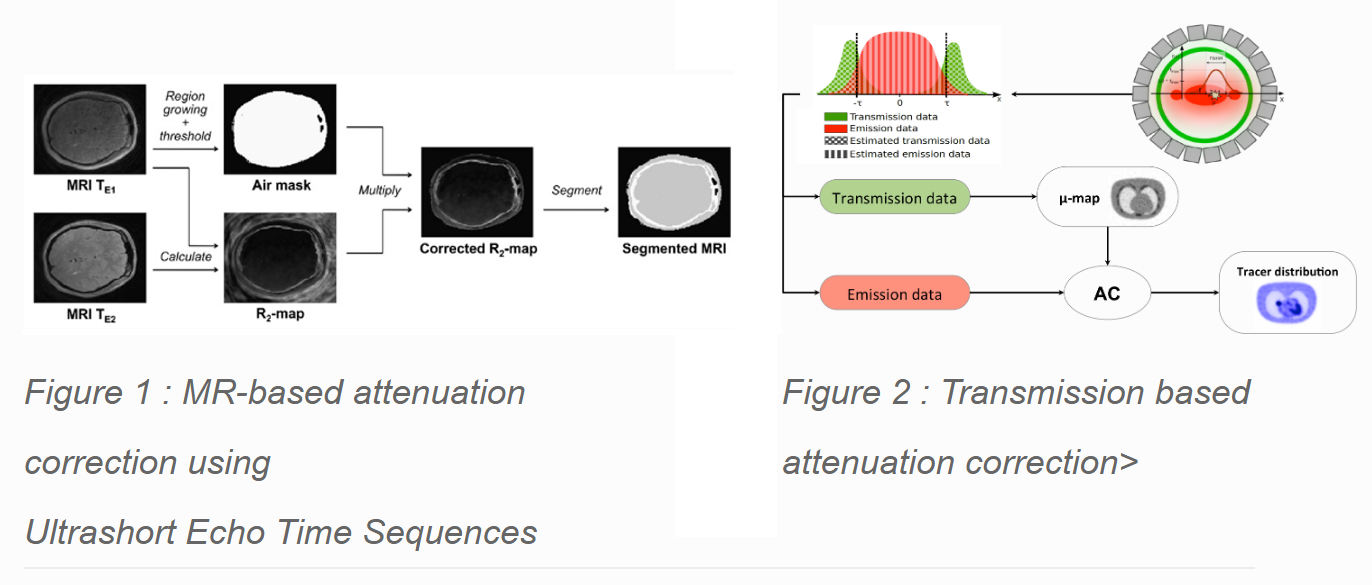
Accurate attenuation correction is necessary to obtain quantitative PET images. In clinical practice, attenuation maps are usually derived from a low-dose CT scan or a PET transmission scan using a rotating source. In combined PET/MRI these methods will most likely not be used. In our group two approaches are investigated to derive the attenuation coefficient at 511 keV. The first approach uses special MR sequences (UTE) in combination with segmentation techniques to derive an attenuation map. In another approach we try to derive the attenuation map from a transmission scan using an annulus shaped transmission source inside the Field-of-View (FOV) of the PET scanner.
Researchers: Pieter Mollet, Vincent Keereman, Ester D’Hoe.
Quantitative conductivity and permittivity imaging using magnetic resonance electrical properties tomography (MREPT)
The frequency-dependent electrical properties (Conductivity and Permittivity) of biological tissue provide important diagnostic information, e.g., tumor characterization (usually the electrical properties of tumor will be higher than that of normal tissues) and also play an important role in estimating the local Specific Absorption Rate (SAR). Magnetic Resonance Electrical Properties Tomography (MREPT) is a method to map the conductivity and permittivity of the tissue using MRI. RF coil of a standard MR system is sufficient for EPT, whereas other methods uses separate electrodes, currents or RF probes for determining the electrical properties of the tissue. This makes MREPT a very promising technique to obtain the conductivity and permittivity in vivo without the need of additional hardware. We will be investigating the non-invasive measurements of the dielectric parameters using MREPT and the implementation of the sequence on the MRI scanner.
Researcher: Prakash Vasudevan
Quantitative imaging for automated brain tumour diagnosis
In Belgium, 800 people are diagnosed with a primary brain tumour each year. Their life expectancy depends strongly on the tumour type, and ranges from several decades for low-grade tumours to only about one year for the most malignant types. An accurate pathological diagnosis is therefore of primary importance. In clinical practice the workflow from initial consultation to therapy involves many steps within a multidisciplinary team of radiologists, neurosurgeons, neuropathologists, nuclear medicine professionals, radiotherapists, etc. In each step of this pipeline, there is a degree of subjectivity involved in the decision process. However, in literature there is growing evidence that a quantitative, and thus objective, approach can yield a correct diagnosis and therapy prediction. The goal of this project is therefore to investigate how a quantitative analysis of medical images (both MRI and PET) can contribute to a personalised medicine. In this project, we will implement a method for automated brain tumour diagnosis, using textural analysis in combination with other quantitative tumour parameters, such as volume, shape and histogram features. Using advanced classification algorithms, such as Support Vector Machines or Convolutional Neural Networks, an algorithm for automated diagnosis will be implemented.

Researcher: Stijn Bonte
SPECT System Modeling and Simulations
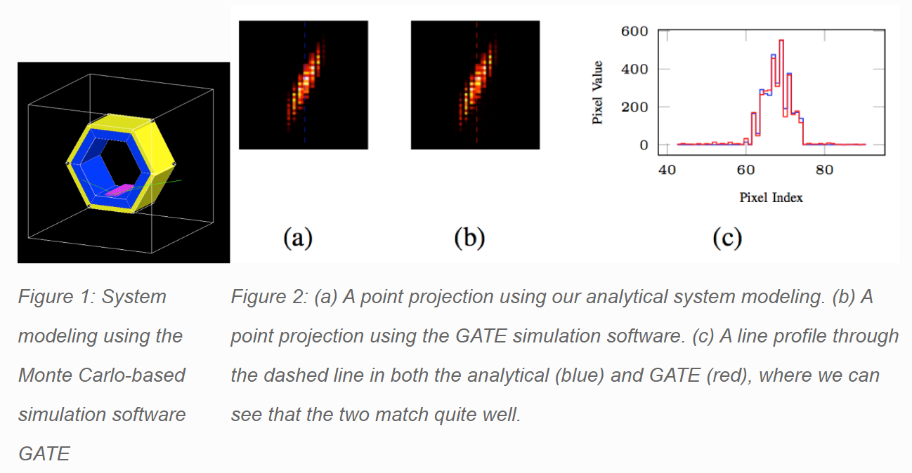
In any Single Photon Emission Computed Tomography (SPECT) scanner, an accurate model of its response is crucial to correctly predict its performance and to get the best reconstruction outcome. This is why we work on improving current methods for analytical modeling of SPECT systems, in particular for the geometrical collimator response in the innovative scanner designs which we develop in our group, which can have quite complex geometries.
On the other hand, we also use statistical software packages (Monte Carlo-based simulation software GATE) that allow us to perform realistic simulations of our system. It is more difficult to use these for the reconstruction itself, but we use them to produce realistic projection data to reconstruct and validate our analytical modeling.
Researcher: Lara Pato
SPECT/MRI compatibility
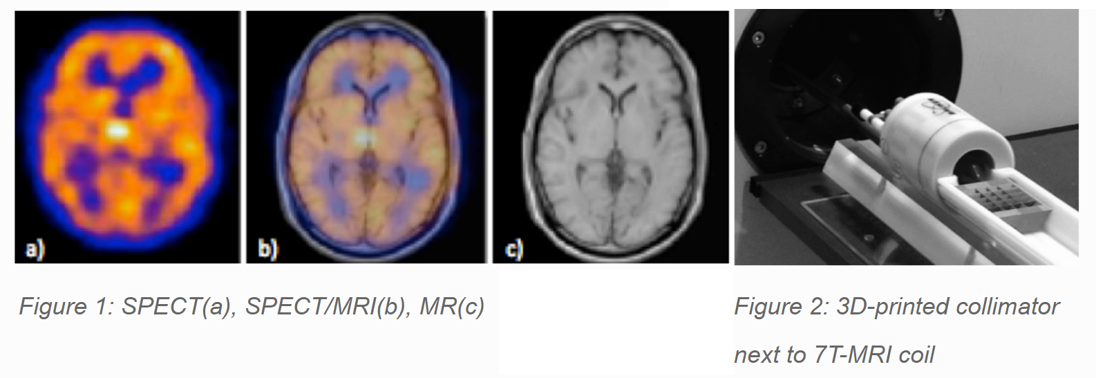
In the last 10 years, there has been a growing interest in multimodality imaging. Integrating different modalities in a single device is advantageous for several reasons: improved patient’s comfort, better diagnosis, and more efficient hospital logistics. Single Photon Emission Tomography (SPECT) and Magnetic Resonance Imaging (MRI) are two complementary modalities: SPECT is a functional imaging technique (Fig. 1a) while MRI is structural (Fig. 1c). A SPECT device consists of two main components: a detector and a collimator. We focus on the MRI-compatibility of SPECT collimators and more specifically 3D-printed tungsten collimators.
Researcher: Karen Van Audenhaege.
The new paediatric TOF-PET: a simulation study
Children are the most challenging and vulnerable nuclear medicine patients due to great differences in their height, weight and physical development. They are more sensitive to consequences of the radiation dose expose than adults and have a higher risk to develop metastatic disease. In order to avoid or at least significantly reduce any risk during examinations, pediatric patients need a suitable diagnostic system that is as fast, as safe and as accurate as possible. Nowadays, there is no a commercial medical imaging modality especially dedicated for children.
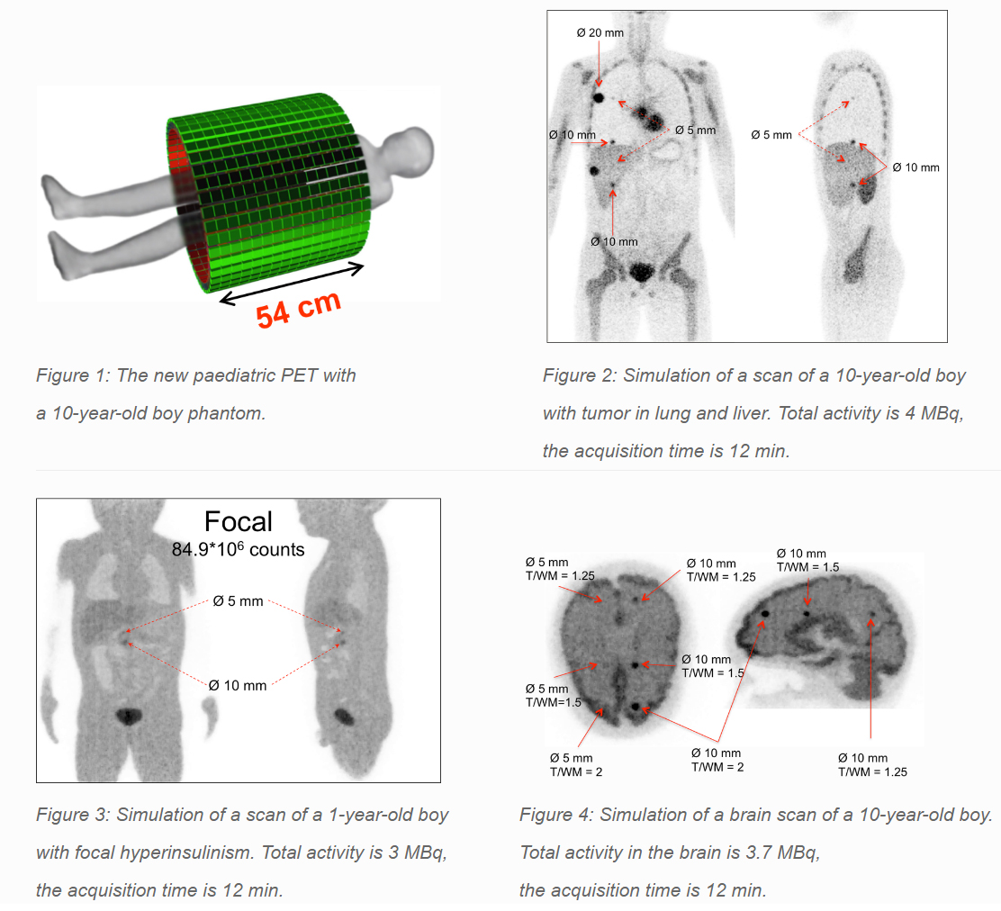
The project aims to evaluate performance of a new pediatric TOF-PET scanner that combines the most recent achievements in radiation detectors, electronics and data processing methods. It is based on monolithic LYSO scintillators coupled to recently developed digital SiPM photodetectors in dual-sided configuration. A distinctive feature of the new pediatric PET is its 150 ps TOF resolution which allows to obtain images of low noise and high contrast. The novel pediatric PET aims to make examinations of children extremely fast, safe and precise.
The figures below show the recent simulation study results performed with voxelized phantoms of children of different age. The results show that the new paediatric PET is able to obtain high quality images in short acquisition times and with ultra-low doses (10-fold reduction).
Researcher: Ekaterina Mikhaylova
Hardware lab
The Infinity hardware lab specializes in the design of SPECT and PET systems. During the last years we have gained experience in radiation detector design, collimator design and system design. The radiation detectors are based on PSPMTs, DSiPMs or analog SiPMs. Custom algorithms have been developed to identify the position of interaction in the detector. We have good contacts with several machining companies to produce SPECT collimators. For this we can choose from traditional techniques (milling, drilling) or additive manufacturing. In-house developed algorithms are used for system calibration and for 3D image reconstruction.
Current research
Past research
- Performance of dSiPMs for compact SPECT scanners
- Performance of dSiPM for PET applications
- SPECT pinhole collimator for optimal detector usage
High resolution monolithic detectors for PET
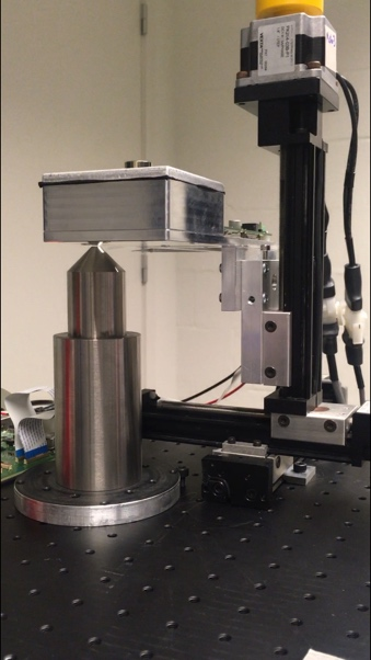
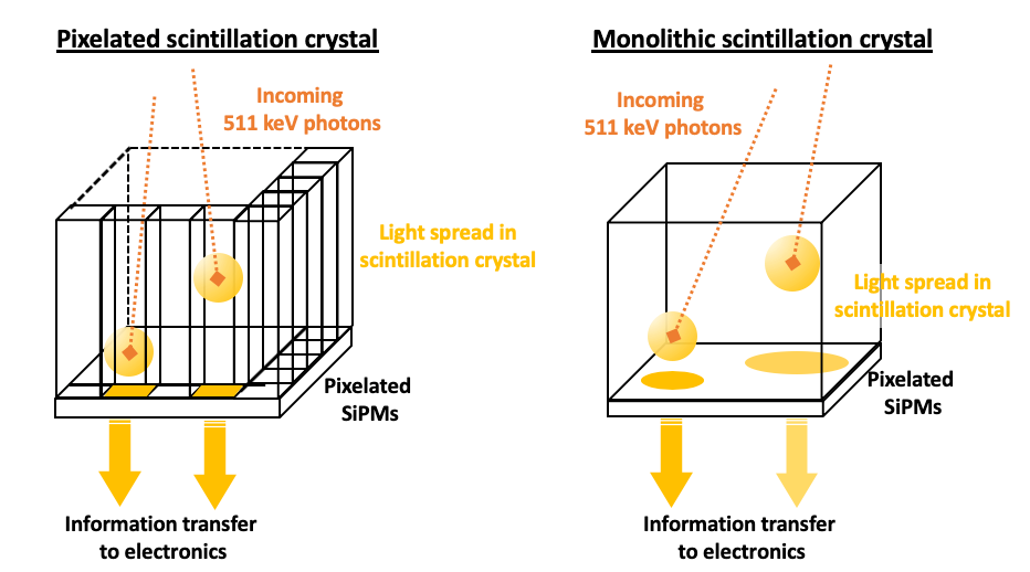
The spatial resolution of positron emission tomography systems is limited by the resolution of the detectors. Clinical PET scanners today employ pixelated detectors based on arrays of narrow and long scintillation crystals, ~ 3x3x18 mm3, coupled to an array of photodetectors, such as silicon photomultipliers. The trend towards smaller scintillation crystal pixels to obtain better spatial resolution negatively affects other desirable parameters like timing and energy resolution, sensitivity, and also cost. For this reason it is likely that future generation PET detectors are based on continuous monolithic scintillation detectors. Monolithic detectors have attractive properties to reach superior 3D spatial resolution while outperforming pixelated detectors in timing, energy resolution and sensitivity. While successfully implemented in preclinical systems, the monolithic detector design has not yet been implemented in any commercial clinical scanner. The aim of this project is the development of a high spatial resolution monolithic detector with application in a clinical PET systems.
Monolithic Time-of-Flight Detectors for PET
The use of (continuous) monolithic crystals has proven to be very successful in preclinical imaging by reaching sub-mm resolution but has not yet been optimised in terms of timing performance in order to do Time-of-Flight PET imaging. In this research we study what the important factors are that affect the time resolution in monolithic scintillators. We look at different readout methods, changing threshold for timestamp determination, different scintillator characteristics etc. The aim of this research is to develop a monolithic PET detector with TOF capabilities that could be implemented in a clinical total-body PET system with high sensitivity and resolution.
Contact: Charlotte Thyssen
Performance of dSiPMs for compact SPECT scanners
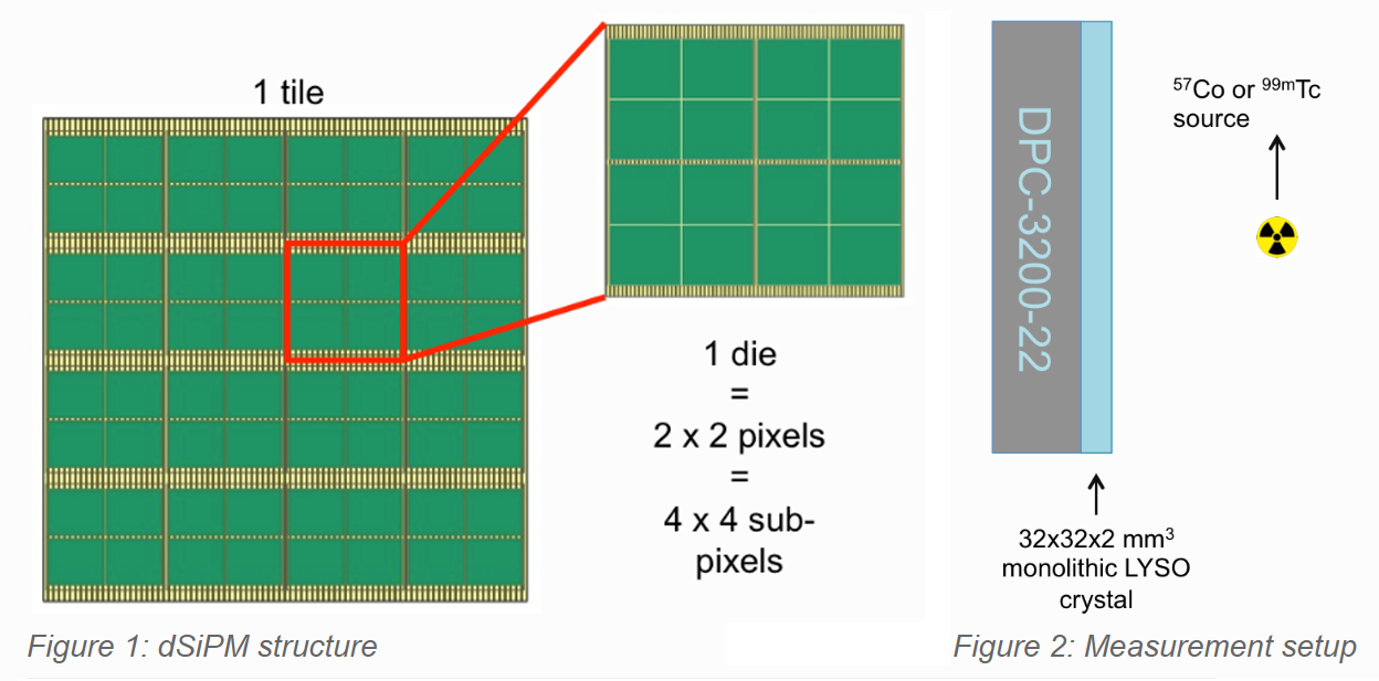
Most preclinical single photon emission computed tomography (SPECT) scanners are based on clinical detectors, resulting in systems with a large footprint. Recently, Philips Digital Photon Counting (PDPC) has introduced a compact, fully digital detector based on solid state technologies, the digital silicon photomultiplier (dSiPM). We are investigating the use of these dSiPMs in combination with thin, monolithic LYSO scintillator crystals as single photon detectors for the development of a compact small animal SPECT scanner.
Researcher: Carmen Bouckaert.
Performance of dSiPM for PET applications
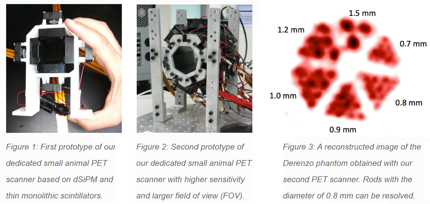
Digital silicon photomultipliers (dSiPM) are solid-state single-photon sensitive devices made of arrays of Geiger-mode avalanche photodiodes (SPAD), in which each SPAD is integrated with CMOS logic circuitry. The integrated CMOS logic provides trigger, precise timestamp, programmable control logic and digital readout for every SPAD. These properties make dSiPM a promising photosensor to be used for Positron Emission Tomography (PET); for both clinical and preclinical applications. In our research we investigate the use of dSiPM for developing a new generation of PET detectors.
Our current interest is focused on evaluating the use of the dSiPM for preclinical PET imaging. Through the combination of the dSiPM with thin monolithic scintillation crystals and 3D events positioning algorithms we achieved compact PET detectors with a sub-millimeter spatial resolution suitable for high-resolution preclinical PET imaging systems. Basing on these detectors we have developed so far two prototypes of compact, high-resolution PET scanner dedicated for mice and rats imaging.
Researcher: Radoslaw Marcinkowski
SPECT pinhole collimator for optimal detector usage
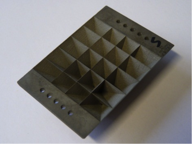
In single-photon emission computed tomography (SPECT), multi-pinhole collimation is used to avoid overlap (multiplexing) of the projections on the detector. However, because of the circular projections of the conventional pinholes the detector surface is not entirely used. We designed a new pinhole geometry called the lofthole, which by its rectangular shape ensures optimal detector usage.
Researcher: Karel Deprez.
Infrastructure
Our research requires state-of-the-art equipment. Below you can find a list of the equipment MEDISIP owns to carry out our experiments. If you have any questions or are interested in using our equipment, please do not hesitate to contact us. You can find our e-mail or phone numbers in the Staff section of this website. Contact persons are Stefaan Vandenberghe, Christian Vanhove and Roel Van Holen.
- BrainAmp MR Plus
- Bruker BioSpin PharmaScan® 70/16
- CT X-CUBE
- IVIS Lumina II
- PET β-CUBE
- U SPECT II
- VEVO 2100
- Xstrahl SARRP
BrainAmp MR Plus

System: BrainAmp MR Plus
Manufacturer: Brain Products
Description: MR compatible EEG system. Enables simultaneous EEG-fMRI recording to investigate the electrophysiological as well as the hemodynamic information of cerebral dynamics.
Bruker BioSpin PharmaScan® 70/16

System: 7 Tesla small animal MRI
Manufacturer: Bruker BioSpin
Description: Up to 50 µm structural resolution for excellent soft-tissue contrast. - rat head / mouse whole body Tx/Rx volume coil (40 mm ID) - rat whole body Tx/Rx volume coil (72 mm ID) - rat brain surface coil (Rx only) - mini flex loop coil (2 cm, Rx only).
CT X-CUBE

System: CT
Manufacturer: Molecubes NV
Description: It allows for fast whole body mouse and rat CT imaging at extremely low dose and excellent soft tissue contrast.
The light weight of this self-shielded imaging unit makes it a truly mobile in-vivo scanner. Advanced workflows, such as gated and dynamic contrast enhanced imaging, can be achieved in a functional and integrated setup. Our iterative reconstruction techniques are available in standard as well as in expert user mode. Intuitive, wireless acquisition software, combined with our multimodal small-animal bed, enables easy and modular multimodal imaging when used along with the β-CUBE (PET).
IVIS Lumina II

System: Optical Imaging
Manufacturer: PerkinElmer
Description: The IVIS Lumina II is capable of imaging both fluorescent and bioluminescent reporters. The system is equipped with up to 21 filter sets that can be used to image reporters that emit from green to near-infrared. An adjustable field of view from 5 – 12.5 cm allows imaging of up to 5 mice or 2 medium size rats. The Lumina II can also accommodate Petri dishes or micro-titer plates for in vitro imaging. The system includes animal handling features such as a heated stage, gas anesthesia connections and ECG monitoring.
PET β-CUBE

System: PET
Manufacturer: Molecubes NV
Description: The β-CUBE is our high-performance preclinical PET imager.
Sub-millimeter image resolution is achieved through the combination of monolithic scintillators, the latest photon counting technology and GPU-based event positioning, and iterative image reconstruction. The 5-ring configuration ensures best-in-class sensitivity over a field of view that is adequate for whole-body mouse and rat imaging at a high count rate. In-house hardware enables dynamic and gated studies. Intuitive, wireless acquisition software, combined with our multimodal small-animal bed, enables easy and modular multimodal imaging when used along with the X-CUBE (CT).
U SPECT II

System: small animal SPECT
Manufacturer: MILabs
Description: Up to 0.35 mm spatial and sub-minute temporal resolution / gated and listmode acquisitions of organs or whole body / high sensitivity: 75 pinholes / collimators: mouse (0.35 and 0.6mm) rat (1mm) dedicated rat brain collimator (0.6mm)
VEVO 2100

System: small animal ultrasound
Manufacturer: VISUALSONICS
Description: 30 µm resolution, up to 1000 fps
Xstrahl SARRP

System: small animal radiation research platform (SARRP)
Manufacturer: Xstrahl
Description: A nozzle-shaped collimator with interchangeable inserts providing circular radiation field sizes of approximately 0.5 and 1 mm in diameter and rectangular fields of 3 x 3, 5 x 5 and 3 x 9 mm for high-precision, small-field, conformal irradiations.
Neuroscience
The neuroscience lab specializes in exploring brain functioning both in small animals as in humans. Multimodal PET, SPECT, EEG, fMRI and optogenetics are used to study dynamic brain functioning in the healthy and epileptic rat brain. In humans, mainly epilepsy is studied and the focus is on EEG data. For this, a close collaboration exists between the neuroscience lab and the neurology department in the University Hospital of Ghent. The neuroscience lab is expert in the field of EEG source imaging (ESI), with advanced forward modelling techniques. Next to this, expertise in brain connectivity analysis is present. Also the use of machine learning in this context is investigated.
Current research
- Combining optogenetics, chemogenetics, intracranial electroencephalography and functional magnetic resonance imaging to investigate abnormal functioning of brain networks during epileptogenesis
- EEG-based brain network analysis of psychosocial stress
- Neuroimaging biomarkers for major depressive disorder: current problems and future directions
Past research
- Advanced forward models for EEG source reconstruction
- Brain-Computer Interfaces
- Seizure onset zone localization from ictal high-density EEG
Combining optogenetics, chemogenetics, intracranial electroencephalography and functional magnetic resonance imaging to investigate abnormal functioning of brain networks during epileptogenesis
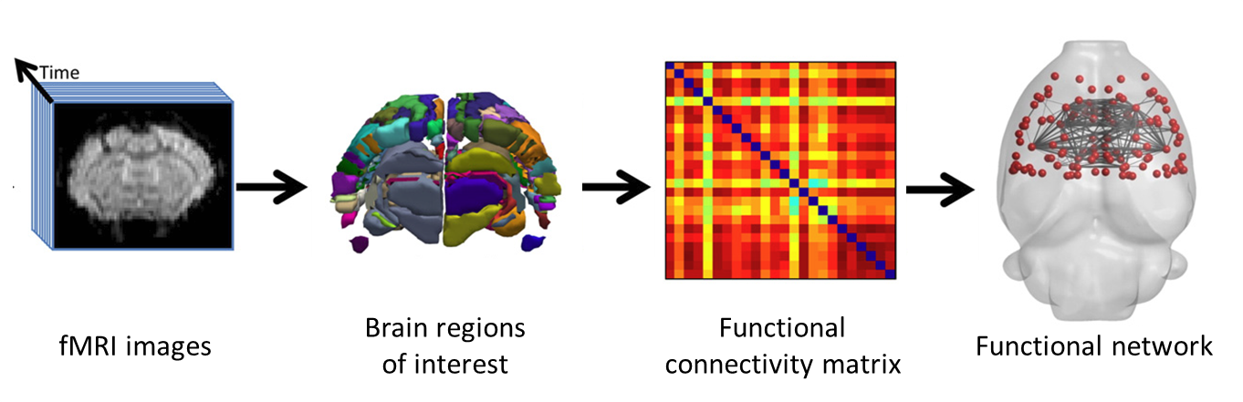
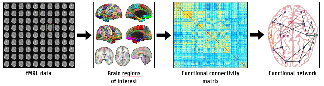
Epilepsy is a disease characterized by recurrent seizures. It is difficult to predict which patients will respond to the different treatment options, because little is known about the development of epilepsy and the neural networks that might be involved. With functional magnetic resonance imaging (fMRI) whole-brain activity can be visualized, functionally connected brain regions can be identified and functional networks can be constructed. These networks can be characterized using graph theory measures. Optogenetics and chemogenetics are new techniques that allow very specific activation or inhibition of neurons, using light and specific drugs respectively. Optogenetics will be used to induce seizures during scanning, while chemogenetics will be used to try to modulate the epileptic network changes and to suppress spontaneous epileptic seizures.
Contact: Emma Christiaen.
EEG-based brain network analysis of psychosocial stress
Psychosocial stress, defined as stress induced by unpredictable, uncontrollable or novel stimuli in a social context, has consistently been linked to a multitude of health complications such as increased risks for heart disease and depression. Three major brain networks, the default mode network (DMN), Salience network (SN) and the central executive network (CEN), are involved in the stress response to psychosocial stress. These networks are defined using fMRI and have been studied extensively, but no EEG-based brain network analyses have been conducted yet. In this project, healthy participants are exposed to both validated as well as novel psychosocial stressors while EEG is collected concurrently, and EEG source modelling is used to investigate how these networks are affected by psychosocial stress, giving novel insights in both within- and between-network interactions related to psychosocial stress exposure. This project is a collaboration between the MEDISIP and Ghent Experimental Psychiatry (GHEP) research groups at Ghent University.

Contact: Gert Vanhollebeke
Neuroimaging biomarkers for major depressive disorder: current problems and future directions
Major depressive disorder, regularly known as depression, is the most common mental health disorder worldwide and is one of the leading contributors to the global burden of disease. Both the prevalence and incidence of major depressive disorder are increasing, showing the need for novel diagnosis and treatment options. In recent years the combination of neuroimaging (EEG and fMRI) and machine learning has been proposed as a solution for this problem, and while initial results show much promise no significant breaktrough has been found yet. This project will firstly investigate the underlying problems in the current approach to machine learning-based diagnosis of depression and secondly propose new methods to combat these problems. This project is a collaboration between the MEDISIP and Ghent Experimental Psychiatry (GHEP) research groups at Ghent University.
Contact: Gert Vanhollebeke
Advanced forward models for EEG source reconstruction
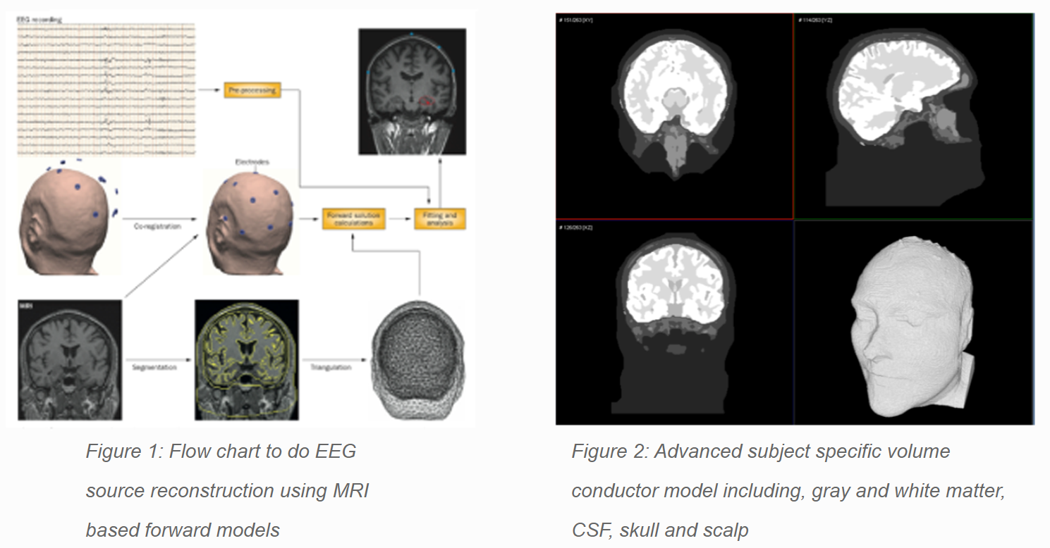
EEG source reconstruction techniques try to reconstruct the generating sources of electrical activity measured on the scalp. In order to do so, a forward model is required. Such a model is characterized by a volume conductor model of the head and a corresponding source space. The volume conductor model reflects the geometrical and electromagnetic properties of the head while the source space constricts the solution space in order to be able to reconstruct the generating sources. We are developing techniques to improve the accuracy of the reconstructed activity using MRI based subject specific finite difference forward models.
Researcher: Gregor Strobbe
Brain-Computer Interfaces
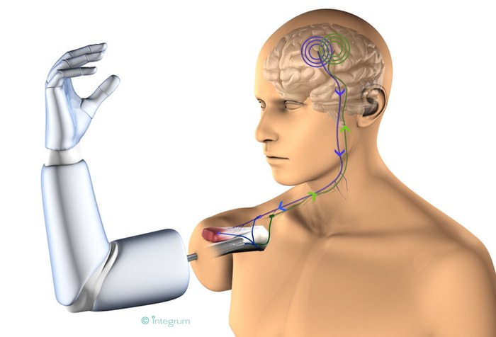
A Brain-Computer Interface (BCI) is a system in which a computer is controlled directly through the use of brain signals, measured as EEG, MEG, fMRI etc. BCIs have a very broad field of application, ranging from computer games to biomedical applications. In this last domain, BCIs are used to increase the standard of living for disabled people. BCIs based on motor imagery enable motor impaired patients to move a wheelchair or an exoskeleton using their brain. Those based on event-related potentials are especially used as communication systems for patiens with the locked-in syndrome (e.g. ALS patients). Extensive research is needed to find accurate methods that extract as much information as possible from the user’s brain waves and transform this information to the right control signals. The final goal of this research is to build accurate, cheap and user-friendly BCIs.
Researcher: Thibault Verhoeven
Seizure onset zone localization from ictal high-density EEG
For patients with refractory epilepsy, surgery is the most efficient treatment option. To make this possible, an accurate delineation of the epileptic focus is necessary. In this research, we investigate how to localize this seizure onset zone based on high-density EEG recordings during a seizure. Therefore, we focus on the fact that there is increasing evidence that epilepsy is a network disease. During a seizure, several brain regions become simultaneously active in an epileptic network and it is hard to discern the main driver of this network. First, we identify the brain activity during a seizure using ESI. Next, we discriminate the epileptic network and try to find it’s driver using functional connectivity analysis.
Researcher: Willeke Staljanssens
Small Animal Imaging
Infinity is the preclinical imaging research facility of Ghent University, providing multimodality-imaging services to national and international customers. It is a consortium of two major research groups: Medisip and the Laboratory for Radiopharmacy. A skilled and experienced scientific staff is running Infinity. Professors Christian Vanhove, Stefaan Vandenberghe, Filip De Vos and Roel Van Holen work together to streamline the research at Infinity. Additionally, professor Christian Vanhove is also responsible for the day-to-day functioning of the lab and the coordination of the partner research, either from within Ghent University, from private companies or from other universities. The major research domains of Infinity are neuromodulation (deep brain stimulation and trans-cranial magnetic stimulation), the use of imaging biomarkers to provide better tumor characterization, stress of the endoplasmatic reticulum (er stress), cardiovascular research to get a better understanding of the formation of aneurysms and plaque, and the development of ‘theranostic’ approaches by combining diagnosis and therapy.
But Infinity is about more than providing service to partners. The lab is actively involved in the multidisciplinary Research Platform on Inflammation and Immunity, called Group-ID. In 5 years time, Group-ID aims to be an international center of excellence in inflammation research, continuously attracting the best students and postdocs, have sufficient independent funding from the government and industry and become a core center of translational research in medicine.
Current research
- Combining optogenetics, chemogenetics, intracranial electroencephalography and functional magnetic resonance imaging to investigate abnormal functioning of brain networks during epileptogenesis
- MR biomarkers for cognitive training in mild traumatic brain injury in rats
Associated research
- Amino-acid PET based radiation treatment in an animal model for glioblastoma.
- Comparison of Ga68-PSMA and F18-PSMA in clinical and preclinical studies.
- Tracing xylem-transported 11C-labelled CO2 in trees: importance and contribution to the carbon metabolism.
Past research
- 18F and 99mTc labeled bile acid analogues to study (altered) hepatobiliary transporter function
- Continuous infusion micro positron emission tomography
- Development of LAT1-specific 18F amino acid PET tracers for the discrimination between low and high grade glioma
- Discriminating high-grade glioma from radiation necrosis in rats using kinetic modeling
- Effect of Cx-mimetic peptide administration on breast cancer brain metastasis development
- Evaluating bone structure surrounding dental implants
- Fast dynamic SPECT
- In-vitro analysis of extracted teeth
- Molecular MRI in early radiation injury detection
- Neural circuit mapping using simultaneous DBS-fMRI and opto-fMRI
- The impact of accelerated High Frequency repetitive Transcranial Magnetic Stimulation (HF-rTMS) on the serotonergic and dopaminergic system in a rat model of depression
- Visualization of the rat somatosensory network
MR biomarkers for cognitive training in mild traumatic brain injury in rats
Traumatic brain injury (TBI) after a fall or car accident can lead to cognitive impairment which limits patients in everyday life. To better understand the underlying pathophysiology, TBI is induced in rats, and the brain is imaged with diffusion MRI, a technique capable of imaging the neural network. In a follow up study the rats will receive cognitive training, similar to training protocols in human patients in order to image the neuroplasticity of the brain. By fitting the diffusion signal to white matter models the neural network can be revealed, and the defects and recovery can be followed up in time. A complementary histological analysis will correlate the biomarkers found in the MRI experiments to the biological basis.
Contact: Kim Braeckman
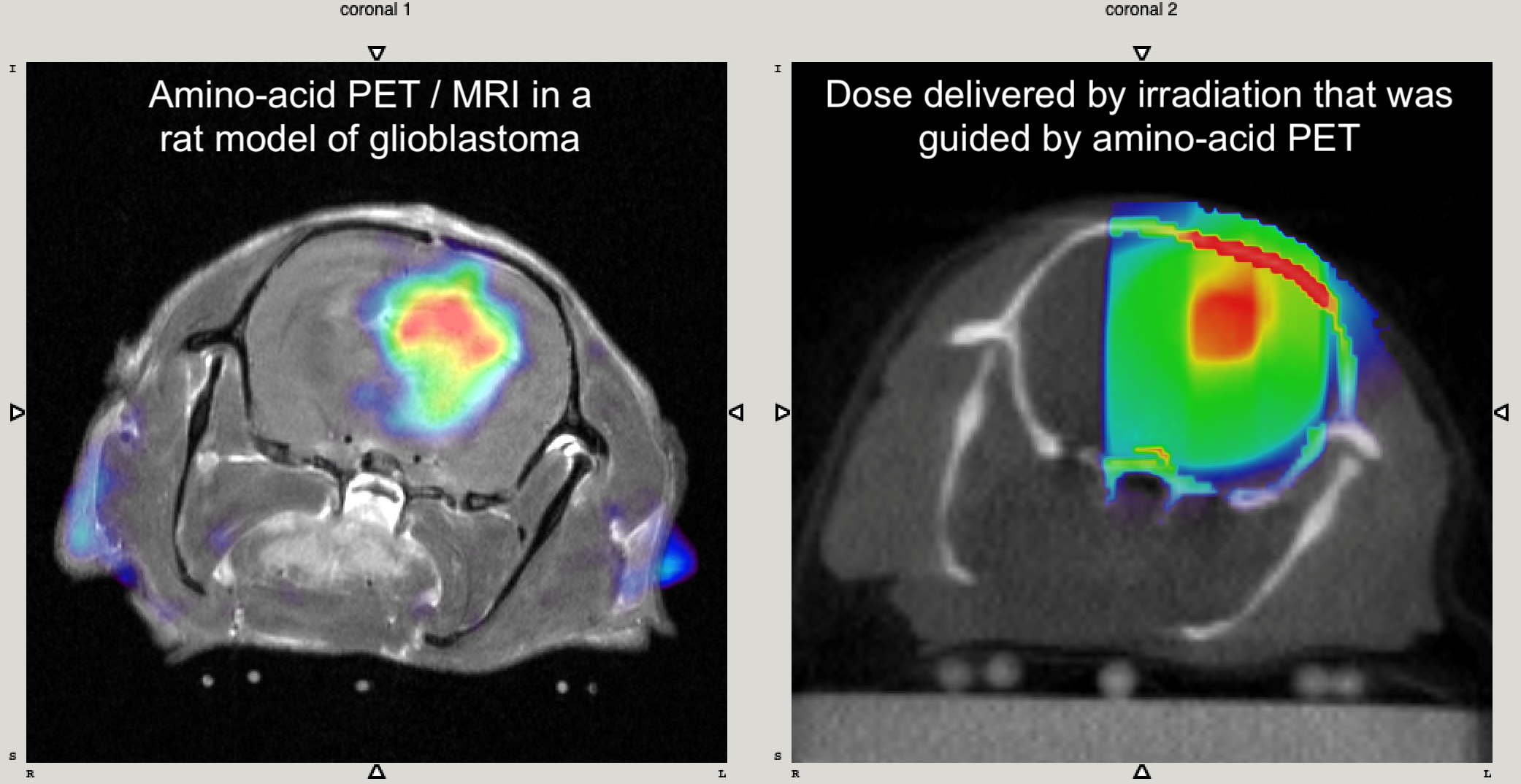
Amino-acid PET based radiation treatment in an animal model for glioblastoma.
Glioblastoma is the most aggressive and most common primary brain tumour in adults. Even with the current state-of- the-art treatment of postoperative combined radiation therapy (RT) and chemotherapy, the median survival is only 12-14 months. Therefore, there is an urgent need for alternative treatment strategies in glioblastoma patients. In our research group, we investigate the added-value of dose painting strategies. More specifically the focus is on incorporating molecular imaging information in the RT plan for glioblastoma with the aim of delivering a non-uniform dose to the so called biological tumour volume. Tumour volume delineation for RT planning in glioblastoma is currently based on anatomical imaging techniques, such as computed tomography (CT) and conventional magnetic resonance imaging (MRI). Importantly, these structural imaging techniques cannot identify the most aggressive and/or radiation resistant parts within the tumour, which are probably responsible for treatment failure and tumour recurrence. Moreover, currently the tumour volume is being irradiated with a homogeneous dose. This is because it is assumed that the tumour consists of tumour cells that share similar behavioural characteristics and are therefore equally malignant and/or radiation resistant or sensitive. This assumption is obviously not correct. Based on molecular imaging techniques, such as positron emission tomography (PET), it is clear that the tumour volume must be considered as heterogeneous regarding its biological features. Using PET-guided dose painting strategies, a non-uniform dose can be delivered to the tumour based on specific biological features. The rationale is that a non-uniform dose distribution with a higher dose to the most active and/or radiation resistant tumour regions, while keeping the total tumour dose constant, could improve local tumour control without adding toxicity to the adjacent brain.
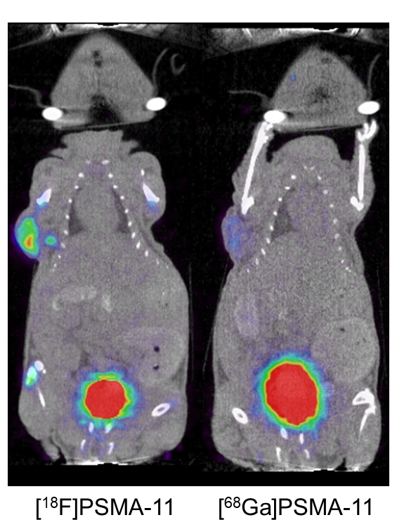
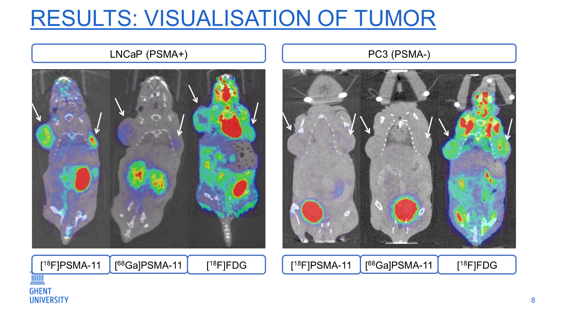
Comparison of Ga68-PSMA and F18-PSMA in clinical and preclinical studies.
Prostate-specific membrane antigen (PSMA) is highly overexpressed in prostate cancer. Many PSMA analog radiotracers for PET/CT prostate cancer staging have been developed, such as 68Ga-PSMA-11. This radiotracer has achieved good results in multiple clinical trials, but because of the superior imaging characteristics of 18F-fluoride, 18F-PSMA-11 was developed. The aim of this study is to compare the prostate‐specific membrane antigen (PSMA)‐targeting characteristics of PSMA‐11, radiolabeled based on chelation of 18F‐AlF, to those of 68Ga‐PSMA‐11 to image PSMA‐expressing xenografts.
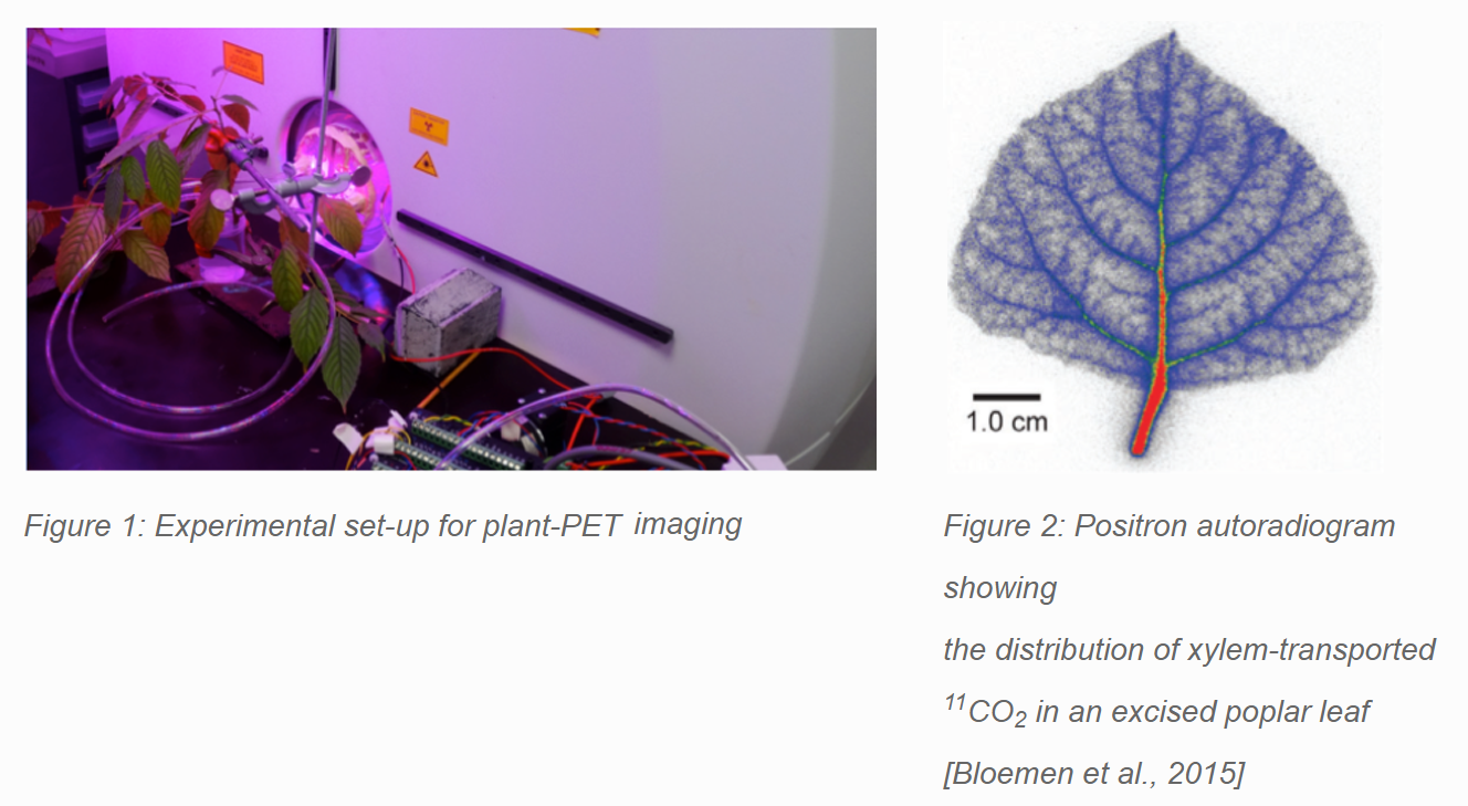
Tracing xylem-transported 11C-labelled CO2 in trees: importance and contribution to the carbon metabolism
Positron-emitting radionuclides like 11C, 13N and 18F are long used in human and animal studies and have led to major breakthroughs. In this respect, positron emission tomography (PET) imaging is receiving increased interest in the field of plant sciences. More specifically, molecules like 11CO2 or 18FDG can, for example, be used as a tool for phenotyping genetically engineered plants and as a new strategy to measure plant responses to drought in a changing climate. The latter being an active and topical research area, yet many questions remain unanswered. In order to understand plant survival and mortality during drought it is crucial to gain more insight into the different mechanisms contributing to both the carbon budget and hydraulic functioning of trees. Hereby, the role of internally transported CO2 in xylem of trees and the associated woody tissue photosynthesis are often overlooked. Our group will focus on these aspects by tracing radioactive 11CO2 in xylem of trees using positron emission tomography (PET) reinforced with magnetic resonance imaging (MRI) for structure-related information. By examining different tree species, subjected towards changing atmospheric conditions, essential insights will be acquired to understand how a given environment will affect plant physiological processes and growth in vivo.
Researcher: Jens Mincke
18F and 99mTc labeled bile acid analogues to study (altered) hepatobiliary transporter function
Numerous drugs are substrate or inhibitor of hepatobiliary transport mechanisms. This can lead to drug induced liver injury. Therefore, it is important to assess these possible interactions from early on during drug development.
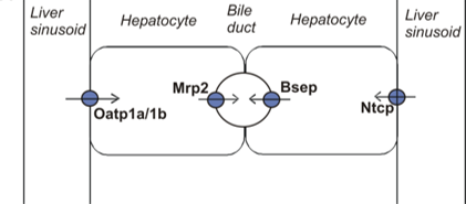
Aim: synthesis of radiolabeled substrates (bile acid analogues) which can be used for molecular SPECT and PET imaging. This is a non-invasive in vivo method to allow visualization and quantification of normal and disturbed hepatobiliary transport. The use of a non-invasive method can significantly reduce the cost for preclinical drug development.
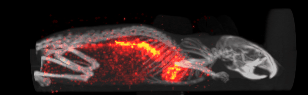
Researchers: Stef De Lombaerde, Sara Neyt
Continuous infusion micro positron emission tomography
Micro Positron Emission Tomography, or µPET, is a functional imaging technique that provides very high sensitivity; ideal to perform dynamic studies. Observing metabolic changes during dynamic µPET is challenging. Injecting a single bolus of the radiotracer implies the need of decay correction. Due to the decrease in radiotracer concentration over time, image quality will decrease in late frames of the dynamic µPET acquisition. By using continuous infusion of the radiotracer subtle or fast changes in the metabolism can be observed, with equal image quality over the complete duration of the dynamic µPET acquisition. Rat brain stimulation experiments have been investigated using this technique.
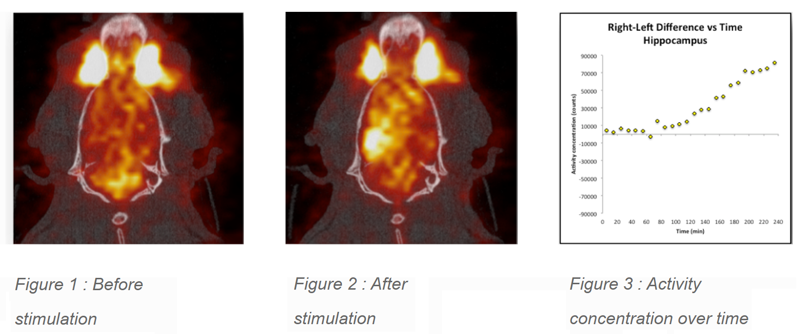
Contact: Chris Vanhove, Vincent Keereman
Development of LAT1-specific 18F amino acid PET tracers for the discrimination between low and high grade glioma
The use of O-(2-[18F]fluoroethyl)-L-tyrosine (18F-FET) as a tumor PET tracer has several limitations including the relatively low affinity for the L-type amino acid transporter (LAT1).
Aim: Synthesis of [18F]-fluoroalkylated phenylalanine analogues with higher affinity for the LAT1 transporter. A LAT1 specific amino acid PET tracer would
allow grading between low and high grade glioma and result in a faster detection of tumor progression.
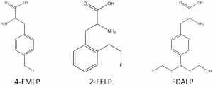
Researcher: Jeroen Verhoeven
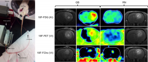
Discriminating high-grade glioma from radiation necrosis in rats using kinetic modeling
Discriminating high-grade glioma from radiation necrosis in rats using kinetic modeling and graphical analysis with three different PET tracers: 18F-fluoromethylcholine (FCho), 18F-fluoroethyltyrosine (FET) and 18F-fluorodeoxyglucose (FDG).
Researcher: Julie Bolcaen.
Effect of Cx-mimetic peptide administration on breast cancer brain metastasis development
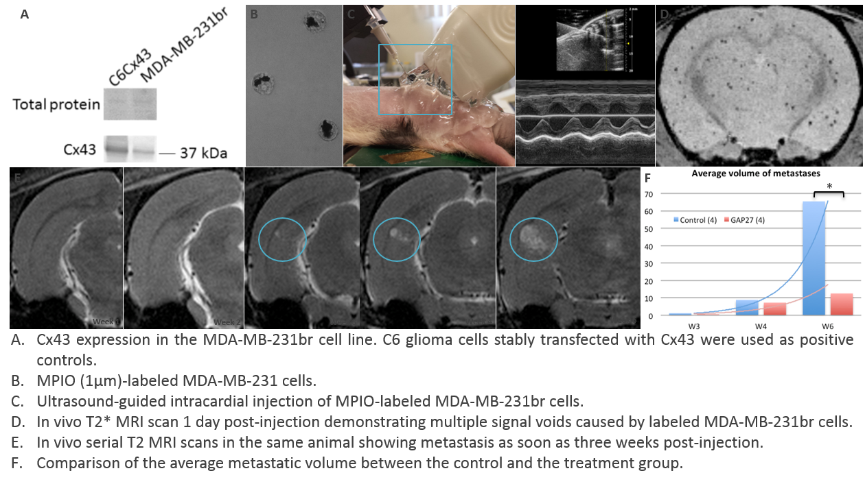
Brain metastasis occurs in 10-15% of the breast cancer patients and entails a strong component of tumor-host interactions. Recent evidence suggests a crucial role for transmembrane connexin (Cx) proteins, primarily Cx43, in the tumor-host interactions by forming intercellular communication pathways. GAP27 is a Cx-mimetic peptide that inhibits Cx-based intercellular communication.
We examine the effect of GAP27 on the initial seeding and subsequent colonization of the brain by using a xenograft model for rat brain metastasis that allows follow-up by molecular magnetic resonance imaging (MRI). The MDA-MB-231br/EGFP breast cancer cell line was used for this purpose.
Researcher: Valerie De Meulenaere.
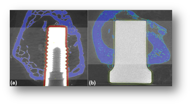
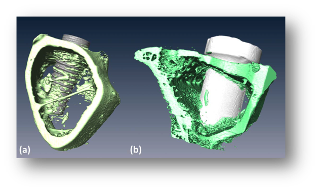
Evaluating bone structure surrounding dental implants
Although histology has proven to be a reliable method to evaluate the ossoeintegration of a dental implant, it is costly, time consuming, destructive, and limited to one or few sections. Micro Computed Tomography, or µCT, is fast and delivers three-dimensional information, but this technique has not been widely used and validated for histomorphometric parameters yet. This study compared µCT and histomorphometry by means of evaluating their accuracy in determining the bone response to two different implant materials.
Contact: Chris Vanhove
Fast dynamic SPECT
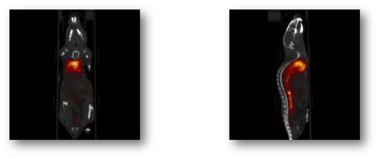
In-vivo micro Single Photon Emission Computed Tomography, or µSPECT, is a functional imaging technique that can provide sub-mm spatial resolution by using pinhole technology. The µSPECT system installed at Infinity (MILabs U-SPECT-II) uses 75 pinholes focused on the same part of the field-of-view, resulting in sub-mm with relatively high sensitivity. This allows for fast dynamic µSPECT to visualize for example hepatobiliary transport using Tc-99m labeled mebrofenin.
Contact: Chris Vanhove
In-vitro analysis of extracted teeth
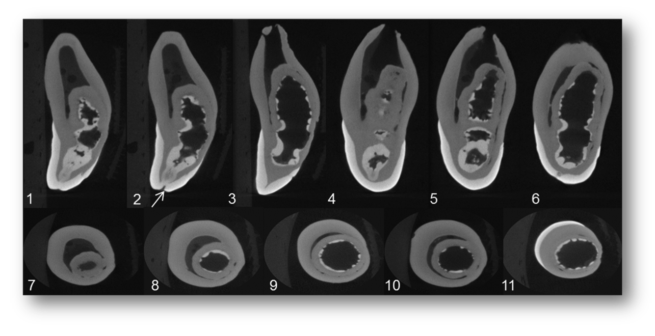
Micro Computed Tomography, or µCT, is an anatomical imaging technique that can provide three-dimensional images at very high spatial resolution. It is an imaging technology that can help in precise, highly accurate, and non-destructive in vitro analysis of teeth.
Contact: Christian Vanhove
Molecular MRI in early radiation injury detection
With conventional magnetic resonance imaging, radiation injury in healthy tissue is only visible at a late stage. Its sensitivity is too small to allow molecular imaging. However, labeling of superparamagnetic particles, e.g., micron-sized particles of iron oxide (MPIOs) with highly specific antibodies, can circumvent this issue. Using this molecular MRI technique, we investigate the early signs of radiation injury, focusing on inflammation-related intravascular molecules.

Contact: Benedicte Descamps
Neural circuit mapping using simultaneous DBS-fMRI and opto-fMRI
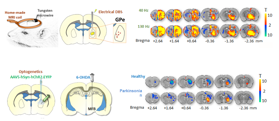
Optogenetics is a new method that uses a combination of optical and gene technologies to manipulate a specific group of neurons with an unprecedented spatial, temporal and cellular specificity. This is reflected in our results since the fMRI-response maps demonstrate a far more selective response to light stimulation as opposed to electrical stimulation (DBS = Deep Brain Stimulation or electrical stimulation; opto = optogenics or light stimulation).
Researcher: Nathalie Van Den Berge.
The impact of accelerated High Frequency repetitive Transcranial Magnetic Stimulation (HF-rTMS) on the serotonergic and dopaminergic system in a rat model of depression
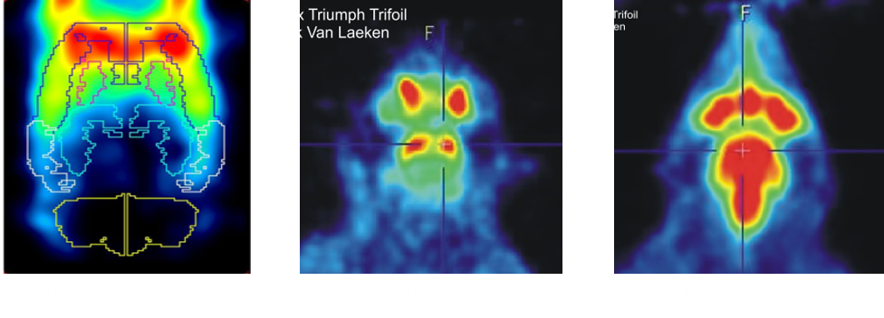
Accelerated HF-rTMS targeted to the DLPFC is a new therapeutic strategy that has shown to be successful in the treatment of major depression in humans. It makes use of an intensified rTMS protocol where both the number of pulses/session and the number of daily sessions have been drastically increased compared to conventional rTMS protocols. Hereby the treatment duration is shortened from several weeks to only four days and beneficial clinical results are obtained much faster. Despite its usefulness, up to now little is known about the basic molecular mechanisms that support these therapeutic effects.
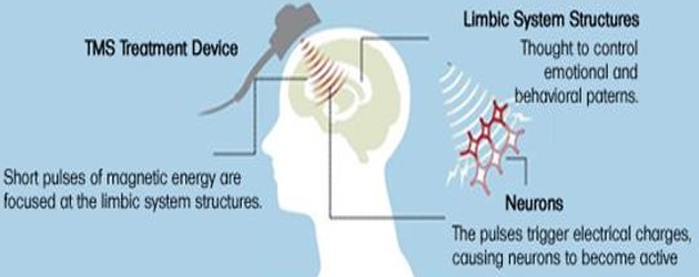
By using the PET-radiotracers [18F]altanserin, [11C]raclopride and [11C]DASB, this study tries to investigate the impact of accelerated HF-rTMS on respectively the availability of the serotonin 5-HT2a receptor, the dopamine D2 receptor and the serotonin transporter in a rat model of depression. Imaging these neurotransmitter systems, which both play an important role in the pathology of depression, can make a substantial contribution to unravel the neurological mechanisms involved in (accelerated) HF-rTMS and to further optimize the stimulation protocol.
Researchers: Nick Van Laeken, Glenn Pauwelyn
Visualization of the rat somatosensory network
Functional magnetic resonance imaging, or fMRI, is an imaging technique to investigate neuronal activation. It is well known that non-noxious electrical stimulation of the rat forepaw leads to neuronal activation of the somatosensory network. Neuroimaging studies have been performed to visualize the entire rodent somatosensory network, especially the subcortical regions. A data-driven technique (Independent Component Analysis, ICA) was used to identify the somatosensory network.
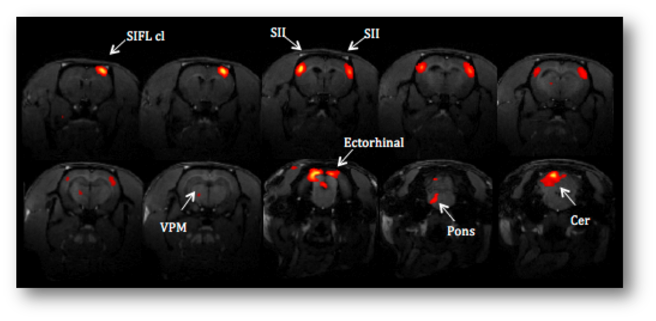
Contact: Chris Vanhove
Collaborations
External non-academic
- partners Barco N.V., Belgium
- CERN European Organization for Nuclear Research, Switzerland
- Fondazione Bruno Kessler, Italy
- GE Medical systems SCS, France
- Inside Matters, Belgium
- Layerwise, Belgium
- MR solutions Ltd., United Kingdom
- Philips Digital Photon Counting, Germany
- VIB, Belgium
External academic partners
- Institut Jules Bordet, Department of Nuclear Medicine, Belgium
- Katholieke Universiteit Leuven, Belgium
- King’s College London, Imaging Sciences and Biomedical Engineering, United Kingdom
- Maastro Clinic, the Netherlands
- Mount Sinai School of Medicine, Translational and Molecular Imaging Institute, USA
- Technische Universiteit Delft, the Netherlands
- Universidad de Antioquia, Colombia
- Universitair Medisch Centrum Leiden, the Netherlands
- Universitair Ziekenhuis Brussel, Belgium
- University of Arizona, Center of Gamma-Ray Imaging and the College of Optical Science, USA
- University of Geneva, EEG and Epilepsy Unit, Switzerland
- University of Heidelberg, Germany
- University of Massachusetts Medical School, USA
- University of Pennsylvania, Physics and Instrumentation Group, USA UT Health Science Center, San Antonio, Texas, USA
- Vrije Universiteit Brussel, Belgium
Internal partners
- Universiteit Gent, vakgroep INTEC-IBCN
- Universiteit Gent, vakgroep Inwendige Ziekten – Laboratorium voor Klinische en Experimentele Neurofysiologie
- Universiteit Gent, vakgroep Sport- en Bewegingswetenschappen
- Universiteit Gent, vakgroep ELIS – Data Science Lab
- Universiteit Gent, vakgroep ELIS – MMLAB
- Universiteit Gent, vakgroep Experimenteel-Klinische en Gezondheidspsychologie
- Universiteit Gent, vakgroep Experimentele Psychologie
- Universiteit Gent, vakgroep INTEC-WICA
- Universiteit Gent, vakgroep Radiologie en Nucleaire Geneeskunde
- Universiteit Gent, vakgroep Farmaceutische Analyse – Laboratorium voor Radiofarmacie
- Universiteit Gent, vakgroep Spraak-, Taal- en Gehoorwetenschappen
- UZ Gent, dienst Neurologie
- UZ Gent, dienst Nucleaire Geneeskunde
Innovation & spin-offs
Our group is a part of the Institute of Biomedical Technology (IBiTech) and is involved in several projects supported by iMinds, an independent research institute founded by the Flemish government. Medisip is also a member of HyCT (dr. E. Van Steenkiste), a valorization consortium from Ghent University. his consortium constantly monitors the valorization potential of our research. As a result, our research group has filed several patents over the past years.
In January 2016, Molecubes, a spin-off of Medisip, was launched. Molecubes focusses on offering a more easy and flexible approach to in vivo scanning of lab animals in the life science research industry. Building world's smallest preclinical imaging cubes, allowing you to combine SPECT, PET and CT imaging, Molecubes offers a solution that fits into almost any lab space. As such, state-of-the-art imaging technology is brought closer to biologists and researchers.
Another spinoff Epilog was launched later this year. The spinoff creates software to automatically detect epileptic phenomena in EEG recordings and localize their origin in the patients' brain.